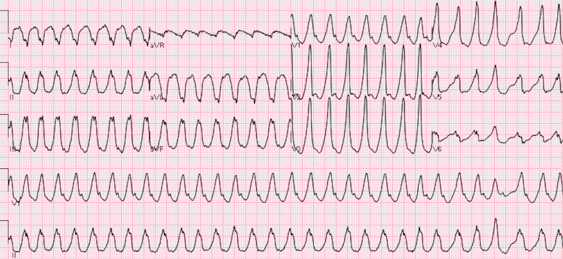
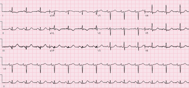
 55 year old male with chief complaint of palpitations. Denies any chest pain, shortness of breath, diaphoresis, or syncope. His past medical history is significant for diastolic congestive heart failure, type 2 diabetes mellitus, hypertension, and hyperlipidemia. Per patient he had a diagnosis of atrial fibrillation vs ventricular tachycardia 2 years prior, but he is unsure of which one exactly.
BP: 153/83 HR: 183 RR: 18 O2 on RA: 99% Temp: 36.3
ECG from triage is shown…
Before reading on, try to come up with your own interpretation of this ECG before moving on to the final impression

- Rate: Ventricular rate 180
- Rhythm: A-V Dissociation
- Axis: Extreme Axis
- QRS: Wide Complex (Consistent with VT)
- Final ECG Interpretation: Wide Complex Tachycardia consistent with VT
When confronted with a 12-lead ECG that shows wide complex tachycardia (WCT), is a correct diagnosis vital for acute treatment, prognosis, and even subsequent work-up? The vast majority of WCT is due to ventricular tachycardia (VT) or supraventricular tachycardia with aberrancy (SVT-A). It is taught that the ECG remains the cornerstone for diagnosis of VT vs SVT-A, and there are several criterion that can be used to help in differentiating WCT. Obviously, a physician cannot memorize all of the algorithms, so if you were to pick one, which would it be? or should you even bother picking one?
What are the criteria used for differentiation of VT vs SVT-A?
- Brugada Algorithm [1]
- Vereckei Algorithm [2]
- R-Wave Peak Time (RWPT) in Lead II [3]
- Griffith Algorithm [4]
- Bayesian Algorithm [5]
- Sasaki Rule (NOT YET VALIDATED)
What are the original article sensitivities, specificities, and accuracies of the above algorithms? [6]

What are the subsequent sensitivities, specificities, and accuracies of the above algorithms? [6]

Case Conclusion: Pt was given 150mg of amiodarone and converted to NSR within 1 minute of receiving this medication (Shown below).

Patient ended up having an extensive workup including:
- Heart catheterization: Minimal coronary artery disease with a 70 – 80% occlusion of a distal obtuse marginal vessel
- Cardiac biopsy: No pathology consistent with amyloidosis, sarcoidosis, or myocarditis
- EP Study: LVOT Tachycardia secondary to aorto-mitral valve continuity
- Final Dx: LVOT Tachycardia
Take Home Points
- In unstable patients, JUST USE ELECTRICITY. Synchronized cardioversion works no matter what the diagnosis is.
- Most of the algorithms are not as sensitive, specific, or accurate as their original studies
- All algorithms have only moderate accuracy and should NOT be used in determining Vtach vs SVT with aberrancy, just assume Vtach!!
References:
- Brugada P et al. A New Approach to the Differential Diagnosis of a Regular Tachycardia with a Wide QRS Complex. Circ 1991. PMID: 2022022
- Vereckei A et al. New Algorithm Using Only Lead aVR for Differential Diagnosis of Wide QRS Complex Tachycardia. Heart Rhythm 2008. PMID: 18180024
- Pava LF et al. R-Wave Peak Time at DII: A New Criterion for Differentiating Between Wide Complex QRS Tachycardias. Heart Rhythm 2010. PMID: 20215043
- Grffith MJ et al. Ventricular Tachycardia as Default Diagnosis in Broad Complex Tachycardia. Lancet 1994. PMID: 7905552
- Lau EW et al. The Bayesian Approach Improves the Electrocardiographic Diagnosis of Broad Complex Tachycardia. Pacing Clin Electrophysiol 2000. PMID: 11060873
- Jastrzebski M et al. Comparison of Five Electrocardiographic Methods for Differentiation of Wide QRS-Complex Tachycardias. Europace 2012. PMID: 22333239


